microscope

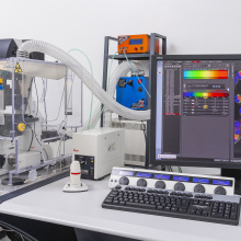
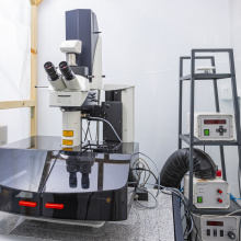
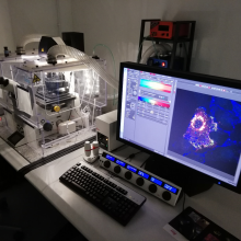
NIKON PCM2000 confocal scanning microscope

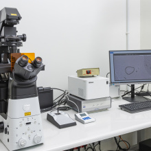
The microscope is equipped with three lasers with wavelengths 488, 546 and 633nm, a HBO mercury lamp and an ultra-sensitive Hamamatsu Orca C4742 color camera. The system allows you to collect images with very low fluorescence intensity. Available lenses: 4x / 0.13, 10x / 0.30, 20x / 0.45, 40x / 060, 63x / 1.4 oil immersion, 100x / 1.4 oil immersion. It enables imaging in bright field, phase and Nomarski contrast, in superfluous light and fluorescence.

Scanning confocal microscope LEICA SP8X with white laser

The white laser enables the selection of any wavelength of the excitation light with an accuracy of 1 nm in the range from 470 nm to 670 nm. Additionally, the microscope is equipped with an argon laser with five excitation lines (458, 476, 488, 496, 514 nm) and a 405 nm diode laser. As a result, the system can be fully adapted to the exact excitation parameters of the various fluorescent dyes. The unique AOBS system is responsible for separating the excitation light beam from that emitted in the microscope.

Scanning confocal microscope Leica HCS LSI

Confocal microscope designed for viewing 'large' specimens. The maximum field of view is 484mm2, it is possible to combine images. The microscope is equipped with four diode lasers (405, 488, 561, 635nm) for confocal operation, a white light source (EPI) and a UV light source. While working in the confocal mode, the image is recorded by a single spectral detector with a working range of 430-750nm and spectral resolution of 5nm. In the operating mode of the stereomicroscope, the image can be viewed directly by the operator or using a monochrome CCD camera.