fluorescence

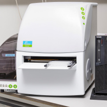
Capillary electrophoresis Sciex P/ACE MDQ Plus with UV and LIF detection

Capillary electrophoresis system equipped with DAD and LIF detectors. System suitable for multiple electromigration techniques implementation (CZE, MEKC, ITP, cIEF, CGE). Typical applications: quality control of pharmaceutical products, chiral analysis, biomolecules analysis. In the lab it is also used for the quality control of the isolates of extracellular vesicles.
https://sciex.com/products/capillary-electrophoresis/p-ace-mdq-plus

2104 EnVision multilabel plate reader

It is a fast and sensitive reader that combines the use of optical filters and two monochromators. It comes with application-specific optical mirror modules, a xenon flash lamp and two high-speed detectors. EnVision also has a standard module for measurements in AlphaScreen ™ technology (PerkinElmer), temperature control, code reader, dispenser and 13 optical filters. The instrument has a 1- to 384-well plate format, scans the cell test well area, and has the ability to read fluorescence from the bottom of the plate.

Scanning confocal microscope LEICA SP8X with white laser

The white laser enables the selection of any wavelength of the excitation light with an accuracy of 1 nm in the range from 470 nm to 670 nm. Additionally, the microscope is equipped with an argon laser with five excitation lines (458, 476, 488, 496, 514 nm) and a 405 nm diode laser. As a result, the system can be fully adapted to the exact excitation parameters of the various fluorescent dyes. The unique AOBS system is responsible for separating the excitation light beam from that emitted in the microscope.

Scanning confocal microscope Leica HCS LSI

Confocal microscope designed for viewing 'large' specimens. The maximum field of view is 484mm2, it is possible to combine images. The microscope is equipped with four diode lasers (405, 488, 561, 635nm) for confocal operation, a white light source (EPI) and a UV light source. While working in the confocal mode, the image is recorded by a single spectral detector with a working range of 430-750nm and spectral resolution of 5nm. In the operating mode of the stereomicroscope, the image can be viewed directly by the operator or using a monochrome CCD camera.